
Nos équipes en France et en Chine vous disent un grand MERCI
Des technologies avancées
pour des tests
cosmétiques efficaces

VENEZ TESTER DES PRODUITS COSMÉTIQUES DE QUALITÉ
- Tests indemnisés
- Sous le contrôle d’un dermatologue
- Produits cosmétiques hauts de gamme
36
17 collaborateurs français
et 19 collaborateurs
chinois
+6000
produits testés
pour de grands groupes
cosmétiques mondiaux
+3000
volontaires déjà inscrits pour
tester des produits cosmétiques
avec nos équipes
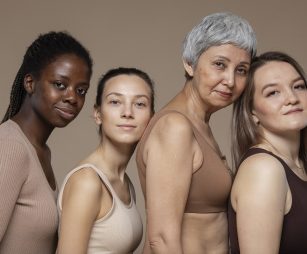
Une présence internationale
Deux centres de recherche :
- COSDERMA France – Bordeaux
- COSDERMA Chine – Wuhan
COSDERMA réalise des recherches biomédicales in vivo sur volontaires sains pour évaluer la Tolérance et l’Efficacité des produits cosmétiques, cosméto-textiles, compléments alimentaires, produits dermo-cosmétiques ou dispositifs médicaux.
Notre savoir-faire dans l’analyse et la conduite d’études cliniques interventionnelles et non-interventionnelles nous permettent de répondre à vos besoins en conformité avec la réglementation en vigueur.
Notre expertise en efficacité et notre niveau d’équipement unique sont reconnus par l’industrie et nous permettent de vous proposer des protocoles innovants et adaptés à vos revendications.
Découvrir nos centres
Une expertise reconnue
Les + COSDERMA
Découvrir les équipements high-tech du laboratoireL'avis de nos volontaires

Une expérience enrichissante
Je participe régulièrement aux études que me propose COSDERMA. Les produits testés sont de qualité. J’apprécie particulièrement les rencontres et les échanges entre femmes lorsque je viens au Centre. Je me sens valorisée avec en plus une rémunération à la clé.
- Maryse, 64 ans,

Fidèle aux tests COSDERMA
Je recommande les tests COSDERMA ! C’est l’occasion de tester des produits de qualité en avant première. L’accompagnement est fait par des professionnels et je n’ai jamais eu de problèmes avec les produits testés.
- Sana, 31 ans,
Découvrez nos dernières annonces

Soin visage
Test indemnisé
entre 50€ et 200€
Voir les annonces
COSDERMA vous accueille toute l’année à Bordeaux pour tester de nouveaux produits cosmétiques haut de gamme (Crème, Shampooing, Anti-rides...)
L'actualité
-
 20 octobre 2025
 -
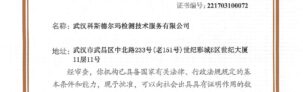
 3 septembre 2024
Nous venons d’obtenir le renouvellement de la norme ISO 9001/V2015 pour la maîtrise de notre… Lire la suite
-
 13 octobre 2022
Notre démarche d’accréditation CMA s’est inscrite dans la continuité des besoins de nos clients, nous… Lire la suite